


Osteochondrosis of the lumbar region is a chronically degenerative dystrophic disease of the lumbar spine, which influences the structure of the intervertebral discs and a number of lumbar vertebrae.It affects people of predominantly working age.It manifests itself in various symptoms, of which the main pain is in the lower back and in the legs and restrict the movements in the lower back.Research methods such as radiography, computer tomography or magnetic resonance imaging of the lumbar spine are used for diagnostics.In this article, you can explain more detailed with the causes, symptoms and methods for diagnosing the osteochondrosis of the lumbar spine.
Osteochondrosis is the result of the body's aging.You can find these or other signs of this disease in almost every person (!) From 25 years.But here is the severity of these changes, the rate of their progress, the degree of clinical manifestations depending on many causes, mainly of how a healthy lifestyle leads a specific person.Moderate physical activity, obligatory morning exercise, the right pose of the body in a series of work (garden, construction, banal cleaning of the house, etc.), the orthopedic mattress are the moments that prevent the development of the osteochondrosis of the lumbar spine.
According to statistics, osteochondrosis of the spine in 80% of cases is the cause of back pain.
How does osteochondrosis develop?
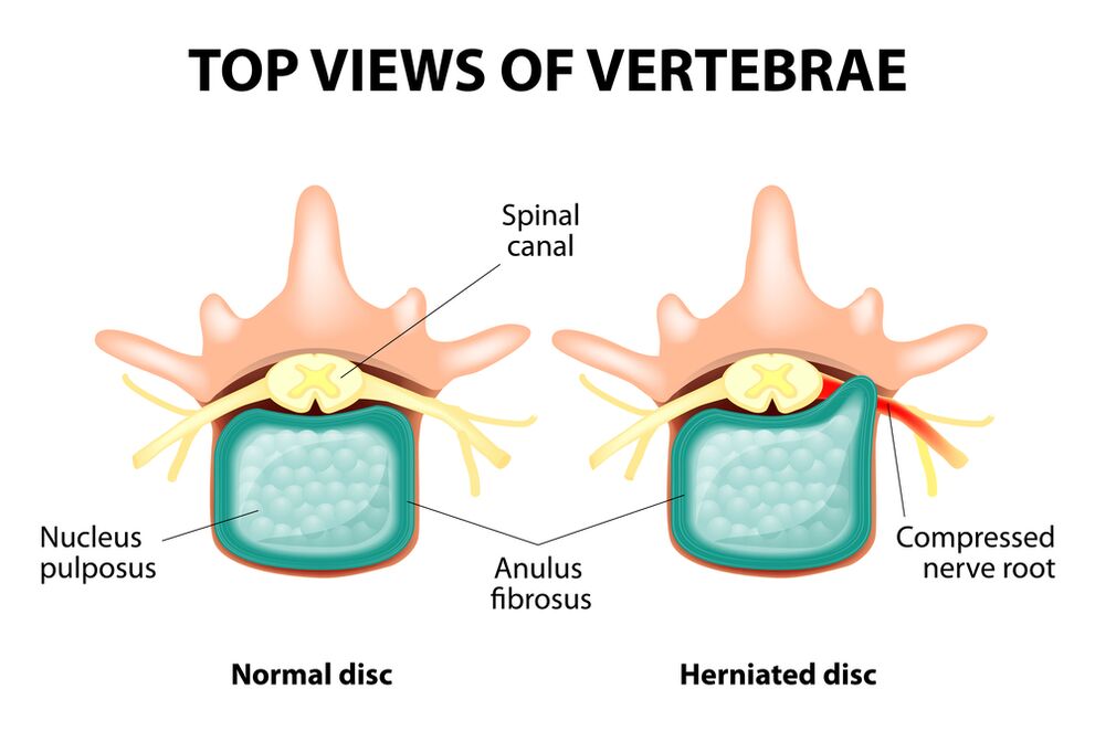
The entire spine consists of separate vertebrae, between the bodies whose intervertebral discs are between hurricanes.This means that there is a hard drive between the two vertebrae.The disc consists of a gallery star (pulpic) core and a fiber ring.The core contains a lot of water and offers depreciation and flexibility of the spine.The fibrous ring is located along the periphery of the Jack Kerne, as if it were holding it in itself.
With a longer increased load in the bracket, it changes its physiological properties, loses water and drying and finally sequences: the disc is flattened, and the vertebral bodies come close together.Together with such processes, the fibrous ring loses its elasticity in the jacket of the core and speaks under the influence of mechanical stress.This is referred to as a lead.Then the fibrous ring tore, and a gelatinous core falls through the resulting gaps: a hernia of the disc occurs.A diagram of two neighboring vertebrae and a disc between them is called vertebral segment increases excessive mobility, which increases the load in the nearby segments.The overload of the neighboring segments triggers a similar pathological process.These changes are called osteochondrosis.
In order to somehow ensure the stability of the spine, bone growth on the edges of the vertebral body are formed, which increases the area of support.This phenomenon is referred to as spondyloses.Changes in the joints between the vertebrae are referred to as spondylo arthrosis.Usually all three pathologies - osteochondrosis, spondyloses, spondylarthrosis - go nearby.
Reasons
Why does osteochondrosis appear?So far there are several theories of the event:
- Mechanical theory: Perhaps the main reason should be regarded as a regular increased stress on the spine.That is why osteochondrosis is an almost mandatory fate of makers, miners, builders and people of such professions.The occurrence of osteochondrosis of the lumbar region is mainly associated with the inclines and the lifting of the severity, forced an unpleasant work.
- Another factor in development is an incorrect attitude that is in the wrong pose, which is particularly relevant to psychiatric employees.
- Sometimes the role of the hereditary characteristics of the structure of the spine and the nutrition of your individual structures is played.
- Traumatic theory: Every trauma to the spine (even the most important) can start a degenerative process;
- Hormonal metabolic disorders and endocrine diseases can adversely influence the metabolism in the tissues of the spine and contribute to the development of osteochondrosis.
- Age theory implies the natural wear of the windows in the life process.
In any case, only one of these theories can explain the occurrence of osteochondrosis.Several factors are more common at the same time.
Obesity plays an important role in the osteochondrosis of the lumbar spine, since it is an overload for the spine itself.The higher the body mass index (degree of obesity), the more pronounced changes in the spine are usually.Among other reasons that cause the appearance of an osteochondrosis, one can note:
- sitting lifestyle;
- Imorly nutrision (fast food, excess sweetness, semi -fixed products: all of this leads to an imbalance of trace elements) and a lack of liquid;
- Anomalies of the structure of the spine (for example the presence of an additional lumbar vertebra);
- Constant wearing shoes with high mental ones;
- Pregnancy (due to the overload of the lumbar spine);
- Sudden termination of training in people who are working professionally in sports;
- Smoking and alcohol abuse: as factors that accelerate the aging process in the body.
Symptoms
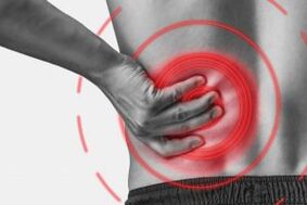
The main manifestation of the osteochondrosis of the lumbar spine is pain.The type of pain, the place of appearance and the distribution gasket depend on which receptors are irritated, ie how large changes in the disc and the surrounding tissues, there is a lead or hernia in which direction the lead was formed.
Reflex and compression syndromes are differentiated by osteochondrosis of the lumbar spine.
Reflex syndromes develop in cases where the receptors of the fibrous ring of the affected disc, ligaments and joint capsules are irritated nearby.They are reflexive, since in addition to pain muscle-tonic, vegetative-vascular or neurodistrophic reflex changes are accompanied, i.e. irritation with reflexes are transferred to other structures, which mainly causes symptoms from the side of the soft tissue.
Compression syndromes occur as a result of the compression (compression) of nerve roots, blood vessels or spinal cord, which are formed by changing osteochondrosis.

Reflex syndromes of the lumbar spine
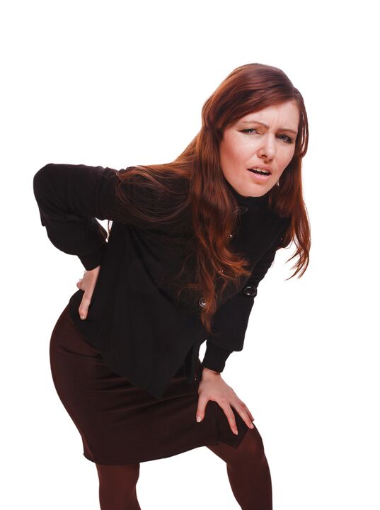
lumbago(Feeling): Acute sudden pain in the lower back, which occurs with an unpleasant movement or at the time of physical tension (much less often - for no apparent reason).It is believed that the appearance of Lumbago is connected to the movement of a coat core within the fibrous ring, i.e. in the early stages of osteochondrosis.The pain is often described as "feeling", "the stake was in the lower back."The patients freeze in the pose in which pain caught them.The slightest movement leads to an increase in pain (sneezing, coughing, an attempt to turn in bed, move your foot).If a person was in an inclined position at the time of the development of Lumbago (which is most common), he cannot straighten up.A pronounced muscle tension in the lumbar spine occurs reflexively.Along the vertebrae in this area, a muscle roller can be felt, which is sometimes visible to the mere eye without touch, and the muscle tension is so pronounced.Feel painful for the patient.Such an increased muscle tone plays an immobilizing role and protects the affected lumbar segment from pathological mobility, which can provoke a deterioration of the state.The natural bend of the spine in the lower back (lordosis) is flattened, possibly the curvature (scoliosis) is possible due to muscle tension.
Lumbargia- Another reflex syndrome of the lumbar strength.This term also means the presence of pain in the lumbar region.In contrast to Lumbago, however, the pain does not occur acute, but gradually within a few hours or even days.The pain is stupid, moderately intensity, intensifies during the movements in a seated or standing position when moving from one position to another.A small relief brings the position of lying down or back with a roller under the lower back, but the passive increase in the limited leg in this position leads to increased pain in the lower back (Lassa symptom).The palpation of the lumbar spine is painful, but the reflex tension of the muscles is less pronounced than with lumbago and sometimes not at all.Movements in the lumbar spine are limited, but possible.This means that the patient can bend to a certain level (and then the pain increases).
sciatica- Another variety of reflex syndrome of the lumbar spine.With this term there is pain in the lower back, which gives the buttocks and legs (on the back).The pain is different, mostly painful, but can regularly intensify by the type of "fireplace" in the leg.Just like lumbargia, it exacerbates movements, walking, efforts, decreasing while lying on the back.The symptom of Lassa is usually positive.The palpation of the lumbar spine is painful and presses a few points (for example in the middle of the line that separates the buttocks from the thigh in the middle of the thigh in the middle of the popliteal fossa).There is a tension of the lower back muscles.The inclinations forward and the sides are limited.
Compression syndromes of the lumbar spine
The clinical feature depends on the structure of compression.
There are nerve roots (vertebral nererves) between the vertebrae in every intervertebral disc: left and right.When pathological formations for osteochondrosis of the lumbar spine (mainly slices of the panes) press the roots, radiculopathy arise, the symptoms of which are different for each root.The increase in pain in the lower back (especially forward), the presence of muscle tension in the lower back, the restriction of movements in the lumbar spine is more common for all radiculopathies in the lumbar region.The following types of radiculopathies of the lumbar spine are most common:
- Radiculopathy L1, L2, L3: Pain occurs in the lower back and conveys the expected thigh.In the same area, the occurrence of paraesthesia (a feeling of crawling, numbness) is possible, disturbing the superficial sensitivity (an acute note from the usual, the feeling of the cold and hot) is not distinguished.The knee reflex decreases, the weakness of the quadriceps of the thigh is revealed;
- Radiculopathy L4: The pain from the lower back results in the front part of the thigh, the inner surface of the knee joint and a little deeper along the inner surface of the lowerbone.Paresthesia can be felt in the same areas, and surface sensitivity is lost (reduced).The weakness in the quadriceps muscle of the thigh also arises, the knee reflex decreases;
- Radiculopathy L5: One of the frequent localizations.The pain gives the buttocks along the outer edge of the thigh along the front surface of the lower leg to the inner edge of the foot and the thumb.Paresthesia can be felt here, superficial sensitivity is disturbed, and here a pain is given here when sneezing and coughing.In addition, there is a difficulty in expanding the foot daring, since the muscle that performs this action is innocent by the kine L5.It is sometimes difficult to stand on a heel with a exposed foot;
- S1 radiculopathy is often also found in osteochondrosis of the lumbar spine.The pain gives the buttocks along the outer edge of the thigh on the outer edge of the lower leg to the outside edge of the foot and on the 5th finger with heels.These zones are characterized by a feeling of paraesthesia, a decrease in surface sensitivity.Achilles reflex is reduced.With the damage to this spine, the weakness of the muscles of the lower leg and the footbeat of the foot develops, so that standing and going on the socks is difficult.
The simultaneous development of radiculopathies of several roots is possible, this is particularly characteristic of L5, S1.It happens that a hernia presses several roots.
If the panel remains, it can press the spinal cord.This is only possible if the hernia is located at the upper reference point, since there is no spinal cord underneath the II lende vertebrae (the roots of the spinal cord are exposed to compression and the ponytail syndrome develops).
If the vessels of the lumbar region are exposed to squeezing that run blood flow to the spinal cord, a spine line develops and with longer compression - myelopathy develops in the event of an acute circulatory disorder.Myelopathy manifests itself through a bilateral weakness of the legs of the legs, begins from the foot and gradually continues.The sensitivity in the legs is disturbed, the Achilles reflex is lost and later knees.It is possible to appear urine disorders (more often, "imperative" urge, which requires immediate satisfaction, urinary incontinence).
Diagnostic methods
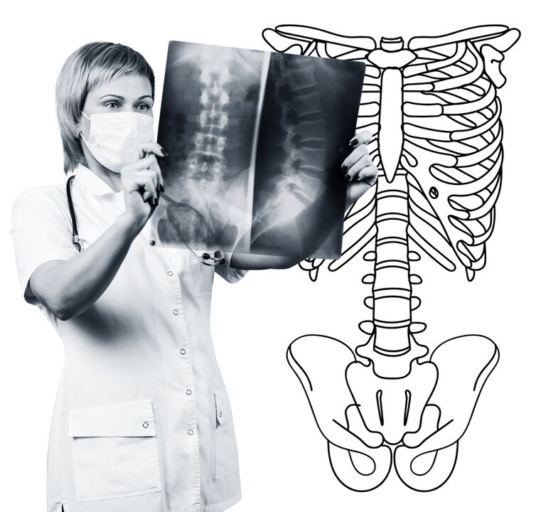
The diagnosis of osteochondrosis of the lumbar spine is based on clinical data and data of additional research methods.The key role is part of methods such as:
- Radiography of the lumbar spine;
- Computer tomography of the lumbar spine;
- Magnetic resonance imaging of the lumbar spine.
The radiography of the lumbar spine is necessarily carried out in 2 mutually vertical projections-the straight and side.Such images enable you to see the shape, contours and structure of the vertebral bodies, the height and shape of the intervertebral discs, the anomalies of the spine and natural bends.Radiograms are generated in sloping projections to display the intermediate vertebral compounds and the grand country.In order to identify the pathological mobility of individual lumbar segments (which is a sign for osteochondrosis), radiography is carried out under the conditions of a functional study, ie in the flexion and expansion of the spine.Usually you can change the height of the intervertebral discs in the front or rear sections in accordance with the direction of the bodies with osteochondrosis due to the functional block of one of the segments neither changes when bending nor when expanding.In the case of pathological mobility, the shift of the vertebrae is determined forward or backwards.The most important x radiation signs of osteochondrosis include the narrowing of the intervertebral slot, the pathological mobility and shift of vertebral bodies, deposition of salts in the disc fabric (calcification), the formation of regional growth of the vertebral body, the compaction of the vertebrae at the border with the affected (subchon -shaped).The radiography of the lumbar spine is a routine of research that gradually loses its importance against the background of the active implementation of new and informative research methods (CT and MRI).The radiography of the lumbar department is now used as a screening diagnostic method.
The CT of the lumbar spine is also carried out using X radiation, but the radial load on the body is much lower than with X -Ray.The study is carried out on the table of a special device - a computer tomography, it is absolutely painless.The resulting images are processed with a computer and enable you to see much more structures than with spinal radiography.
MRI is a method in which electromagnetic radiation is used to create images.The study is also carried out in the position of lying on the table, which calls into the chamber of tomography.MRI is harmless and painless.
With CT or MRI of the lumbar spine you can see all the structures of the spine and carefully examine the intervertebral discs (as well as the coat and the fiber ring) and the intervertebral disc holes, the content of the vertebral canal.Even a minor advantage of the intervertebral disc does not go unnoticed.With these methods (in particular the MRI) you can determine the direction of the window hernia, if available, the degree of compression of nerve roots, spinal cord.Therefore, these research methods are much more informative than radiography when diagnosing an osteochondrosis of the lumbar spine.In addition, you can not only diagnose osteochondrosis, but also other diseases (tumors, circulatory disorders in the spinal cord, abscesses, congenital defects of the structure of the spine and the spinal cord), which is important during the difference diagnosis of pain causes of pain.
Osteochondrosis of the lumbar spine is an illness that most often causes back pain.It is indeed the destruction of the intervertebral discs.Due to the osteochondrosis of the lumbar spine, a person often loses the ability to work because the disease can also lead to pain to violate the mobility of the spine, the inability to sit, stand and go.The symptoms of this disease are not specific and require additional research methods to precisely confirm the diagnosis.The most informative and safest of modern methods for diagnosing osteochondrosis is the MRI of the spine.